Magnetic resonance imaging (MRI) stands as one of modern medicine’s most powerful diagnostic tools, providing physicians with unprecedented views inside the human body. Whether you’re scheduled for your first mri scan or simply curious about this remarkable technology, understanding what an MRI is and how it works can help demystify the experience and highlight why this imaging method has become indispensable in healthcare.
Unlike traditional x ray imaging or ct scanning, mri machines use no ionizing radiation, making them safer for repeated use and particularly valuable for monitoring chronic conditions. This comprehensive guide will walk you through everything you need to know about magnetic resonance imaging, from the basic science behind how it works to what you can expect during your mri exam.
What is MRI?
Magnetic resonance imaging (MRI) is a noninvasive medical imaging technique that uses strong magnetic fields and radio waves to create detailed images of internal body structures. This sophisticated technology harnesses the magnetic properties of hydrogen atoms naturally present in our bodies to generate remarkably clear pictures of soft tissues, organs, and bones.
Mri scanners use magnetic field strengths ranging from 0.2 to 7 Tesla to generate high-resolution images without ionizing radiation. To put this in perspective, most clinical mri machines operate at 1.5 Tesla or 3 Tesla – roughly 30,000 times stronger than Earth’s magnetic field. This powerful magnetic environment allows the scanner to detect minute differences in tissue composition that would be invisible to other imaging methods.
Unlike X-rays and CT scans, MRI does not expose patients to harmful radiation, making it safer for repeated imaging. This advantage proves particularly valuable for cancer treatment monitoring, tracking neurological conditions like multiple sclerosis, and imaging children or pregnant women when medically necessary.
MRI produces cross-sectional images that can be viewed from different angles, similar to slices in a loaf of bread. These detailed pictures provide physicians with three-dimensional insights into anatomical structures and can reveal abnormalities that might be missed by other imaging techniques. The superior soft tissue contrast achieved through magnetic resonance makes MRI the preferred choice for evaluating the brain and spinal cord, detecting ligament injuries, and identifying tumors.
How Does MRI Work?
The physics behind magnetic resonance imaging relies on a fascinating interaction between magnetic fields, radio waves, and the hydrogen atoms that make up roughly 60% of the human body. Understanding this process helps explain why MRI produces detailed images superior to many other imaging methods.
Mri machines create a strong magnetic field that aligns hydrogen atoms in the body in the same direction. Under normal circumstances, these tiny atomic magnets point in random directions. However, when exposed to the scanner’s powerful magnetic field, they align like compass needles pointing north, creating a measurable net magnetization in the tissue.
Radio wave pulses are sent to disturb these aligned atoms, and when turned off, atoms return to position and emit signals. This process, called magnetic resonance, occurs at specific frequencies determined by the magnetic field strength. For hydrogen atoms at 1.5 Tesla, this resonance frequency is approximately 63.9 MHz, while at 3 Tesla it reaches about 127.8 MHz.
A computer converts these signals into detailed images of organs, tissues, and bones. The scanner’s sophisticated software analyzes the timing and intensity of signals returning from different tissues. Since various tissues contain different amounts of water and fat, they emit distinct signal patterns that create contrast in the final images.
Magnetic field gradients help localize signals spatially to create precise images of specific body regions. These gradient coils superimpose additional magnetic fields that slightly alter the resonance frequency of atoms in different locations, allowing the computer to determine exactly where each signal originates within the body.
Types of MRI
Modern mri technology offers various configurations and specialized techniques designed to optimize image quality, patient comfort, and diagnostic capabilities. The choice of mri scanner type depends on the clinical indication, patient needs, and institutional resources.
Open vs Closed MRI
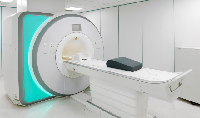
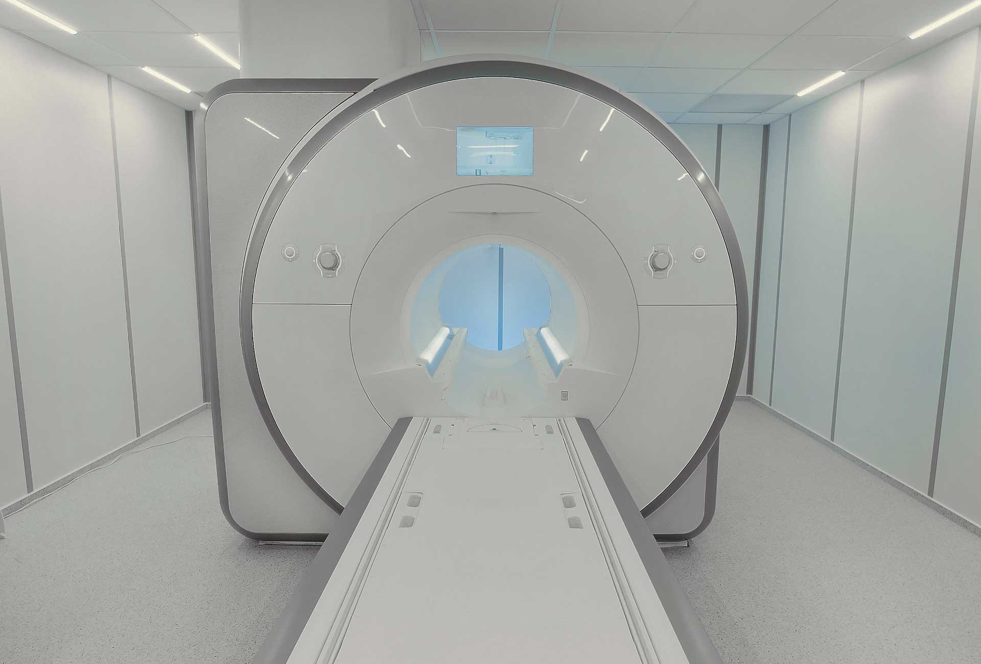
Closed-bore mri machines have a ring of magnets forming a narrow tube, producing the highest quality images. These traditional mri systems feature a cylindrical design with an internal diameter of approximately 60 centimeters. The enclosed magnet configuration provides optimal magnetic field homogeneity and gradient performance, resulting in superior image resolution and signal-to-noise ratio.
 Open mri machines have two flat magnets with space above and below, reducing claustrophobia for anxious patients. This design creates an open environment on the sides, allowing better access for larger patients, children who need parental presence, and individuals who experience severe anxiety in enclosed spaces. Some open systems can accommodate interventional procedures that require physician access during imaging.
Open MRIs produce lower quality images compared to closed-bore machines but offer better patient comfort. The magnetic field strength in open systems typically ranges from 0.2 to 1.0 Tesla, compared to 1.5-3 Tesla in closed systems. This difference affects image resolution and scan time, with open MRI exams often taking longer to complete.
Patients with anxiety can discuss sedation or anesthesia options with their healthcare provider. Many facilities also offer wide-bore scanners as a compromise, featuring closed magnet design with expanded diameter (approximately 70 centimeters) to improve comfort while maintaining high-quality imaging capabilities.
MRI with Contrast
Gadolinium-based contrast material is injected intravenously to enhance image quality and sensitivity for detecting certain abnormalities. These contrast agents, also known as gadolinium containing agents, work differently from the iodinated contrast used in CT scans. Instead of directly blocking radiation, mri contrast agents alter the magnetic properties of water molecules in nearby tissues.
Contrast agents alter magnetic properties of water molecules to improve visualization of certain tissues. Gadolinium shortens the T1 relaxation time of nearby water protons, causing enhanced tissues to appear brighter on T1-weighted images. This enhancement pattern helps radiologists distinguish between healthy tissue and pathologic processes such as tumors, inflammation, or blood-brain barrier disruption.
IV catheter is placed in hand or arm for contrast injection during the procedure. An mri technologist typically establishes intravenous access before the scan begins, using an IV needle to place the catheter. The contrast material is administered through this line during the examination, often after initial non-contrast images have been obtained.
Severe allergic reactions to contrast are very rare but patients should report any known allergies. While gadolinium agents generally cause fewer allergic reactions than iodinated contrast, mild reactions such as nausea, headache, or injection site discomfort can occur. Patients should inform their healthcare provider about any previous adverse reactions to contrast material, kidney problems, or pregnancy status.
Specialized MRI Techniques
Advanced mri techniques extend beyond basic anatomical imaging to provide functional and physiological information that enhances diagnostic capabilities.
Functional magnetic resonance imaging (fMRI) measures blood flow and brain activity to locate critical functions like language and movement. This technique detects changes in blood oxygenation that occur with neural activity, creating maps of brain function that prove invaluable for neurosurgical planning and neuroscience research.
Magnetic resonance angiography (MRA) visualizes blood vessels and flow patterns without requiring invasive catheter procedures. MRA techniques use specialized sequences to highlight moving blood, creating detailed images of arteries and veins throughout the body. This non-invasive approach helps diagnose aneurysms, stenoses, and vascular malformations.
Magnetic resonance elastography (MRE) uses sound waves to assess tissue density and liver disease. By measuring tissue stiffness, MRE can detect liver fibrosis and cirrhosis earlier than conventional imaging methods, providing crucial information for managing liver disease.
Magnetic resonance spectroscopy (MRS) measures metabolite levels in tissues for metabolic disorders. Rather than creating images, MRS analyzes the chemical composition of tissues, detecting molecules like N-acetylaspartate, choline, and lactate that can indicate disease processes or treatment response.
What Does MRI Show?
The versatility of magnetic resonance imaging allows physicians to examine virtually every part of the human body with exceptional detail. The superior soft tissue contrast provided by mri makes it particularly valuable for diagnosing conditions that affect organs, muscles, nerves, and other structures that may appear similar on other imaging tests.
Brain and Spinal Cord Imaging
MRI is the preferred imaging method for neurological conditions, brain tumors, and spinal cord injuries due to its unparalleled ability to distinguish between different types of brain tissue. The brain and spinal cord contain numerous structures with subtle differences in water content and composition that create excellent contrast on mri images.
Functional MRI helps guide brain surgery decisions and assess damage from head injuries by mapping critical brain functions before surgery. Neurosurgeons rely on fMRI to identify areas responsible for language, motor control, and sensory processing, allowing them to plan surgical approaches that preserve essential functions while removing tumors or treating other conditions.
MRI can detect early signs of Alzheimer’s disease and other neurodegenerative conditions through specialized techniques that measure brain volume, detect amyloid plaques, and assess white matter integrity. These capabilities make MRI an important tool for research into dementia and for monitoring disease progression.
Superior contrast between grey and white matter aids in diagnosing CNS disorders such as multiple sclerosis, where the ability to detect subtle changes in brain tissue helps physicians track disease activity and treatment response. Diffusion-weighted imaging can identify acute strokes within minutes of onset, often before symptoms become apparent.
Heart and Blood Vessel Assessment
Cardiac magnetic resonance imaging provides comprehensive evaluation of heart structure and function without exposing patients to ionizing radiation or requiring invasive procedures. This makes it particularly valuable for young patients and those requiring frequent imaging.
Cardiac MRI evaluates heart structure, function, and blood vessel abnormalities with exceptional precision. The technique can measure ventricular volumes, ejection fraction, and wall motion with accuracy that serves as a reference standard for cardiac function assessment. Late gadolinium enhancement imaging can detect myocardial scarring from previous heart attacks or cardiomyopathy.
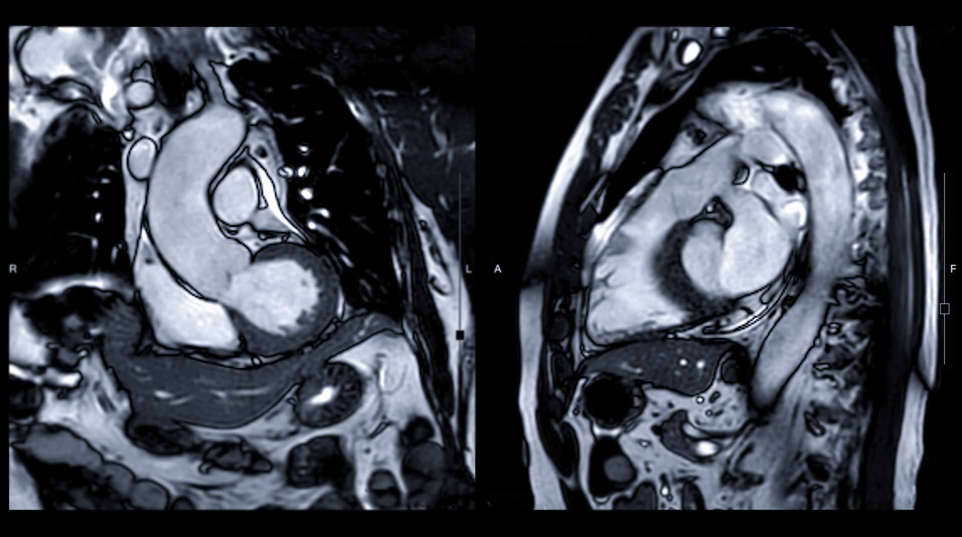
MRI detects cardiovascular conditions without invasive procedures by using specialized sequences that highlight blood flow, vessel walls, and cardiac tissue characteristics. This capability proves especially valuable for diagnosing congenital heart disease, where complex anatomical relationships require detailed visualization.
Provides detailed views of blood flow and vessel integrity for treatment planning through magnetic resonance angiography techniques that can identify aneurysms, stenoses, and vascular malformations throughout the body without contrast injection.
Internal Organ Evaluation
MRI detects tumors and irregularities in liver, kidneys, pancreas, and other organs with sensitivity that often exceeds other imaging modalities. The ability to characterize tissue composition helps distinguish benign from malignant lesions and guides treatment decisions.
Provides detailed soft tissue contrast superior to CT scans for abdominal imaging, particularly in evaluating liver lesions, pancreatic masses, and kidney abnormalities. Multi-parametric liver MRI can detect and characterize focal liver lesions, assess liver fibrosis, and monitor treatment response in cancer patients.
Helps diagnose organ diseases and plan treatments without radiation exposure, making it ideal for young patients and those requiring frequent monitoring. MR cholangiopancreatography (MRCP) provides detailed images of bile ducts and pancreatic ducts without invasive procedures.
Useful for monitoring treatment response in cancer patients through techniques that measure tumor size, blood flow, and cellular density. Advanced sequences can distinguish between viable tumor tissue and treatment-related changes, guiding ongoing therapy decisions.
Musculoskeletal System
MRI evaluates bones, joints, muscles, ligaments, and tendons for injuries and diseases with unmatched soft tissue detail. Unlike X-rays that primarily show bone, mri reveals the complete picture of joint health including cartilage, ligaments, and surrounding soft tissues.
Detects fractures, inflammation, arthritis, and degenerative joint conditions through multiple imaging sequences that highlight different tissue characteristics. T2-weighted images excel at detecting fluid and edema, while T1-weighted images provide excellent anatomical detail.
Provides detailed images of soft tissues around bones not visible on X-rays, making it essential for diagnosing sports injuries like anterior cruciate ligament tears, rotator cuff injuries, and meniscal damage. The ability to visualize cartilage damage helps guide treatment decisions in arthritis management.
Essential for diagnosing sports injuries and planning orthopedic treatments by providing precise anatomical information that influences surgical planning and rehabilitation strategies. MRI can also detect bone marrow abnormalities that may indicate stress fractures, infections, or malignancies.
Breast Imaging
MRI supplements mammography for breast cancer detection in high-risk patients, offering enhanced sensitivity for detecting invasive cancers. Breast MRI uses dynamic contrast enhancement to assess tissue vascularity patterns that may indicate malignancy.
Particularly useful for women with dense breast tissue where mammography may be limited, as MRI is not affected by breast density. This advantage makes breast MRI valuable for screening women with genetic predisposition to breast cancer or strong family histories.
Improves early detection and staging of breast cancer by identifying additional tumor foci that may not be visible on mammography or ultrasound. Pre-operative breast MRI helps surgeons plan treatment approaches and assess for multifocal or multicentric disease.
Helps monitor treatment response and detect recurrence in patients undergoing chemotherapy or following surgical treatment. The high sensitivity of breast MRI makes it an important tool for surveillance in high-risk populations.
MRI Safety and Risks
MRI is generally safe with minimal risks when safety guidelines are followed, but the powerful magnetic field and radiofrequency energy require careful attention to safety protocols. Understanding these considerations helps ensure safe examinations for all patients.
Strong magnetic field can interfere with implanted medical devices like pacemakers and cochlear implants, potentially causing malfunction or unsafe heating. The magnetic field extends beyond the scanner itself, creating zones where ferromagnetic objects can become dangerous projectiles if brought too close to the magnet.
Most modern implants are MRI-safe, but older devices may prevent MRI scans entirely. Patients with metal or electronic devices must undergo careful screening to determine MRI compatibility. Devices may be classified as MRI-safe, MRI-conditional (safe under specific conditions), or MRI-unsafe.
Common implants that may affect MRI safety include:
- Vagus nerve stimulators
- Metallic joint prostheses
- Cochlear implants
- Hearing aids
- Metal implants from previous surgeries
Mild allergic reactions to gadolinium contrast are possible but manageable with medication. While severe reactions are rare, patients should report any history of contrast allergies or kidney disease, as certain gadolinium containing agents may pose risks for dialysis patients or those with severe renal impairment.
Gadolinium-enhanced MRI is usually avoided during pregnancy unless absolutely necessary, as the effects of gadolinium on fetal development are not fully understood. However, non-contrast MRI is generally considered safe during pregnancy when medically indicated.
Tattoos with metal-containing inks may cause discomfort or image artifacts during the scan. Patients should inform the mri technologist about any tattoos, as some pigments contain metallic compounds that can heat during radiofrequency pulse sequences.
A rare but serious illness called nephrogenic systemic fibrosis has been associated with certain gadolinium agents in patients with severe kidney disease. This condition primarily occurs with older linear contrast agents, leading to preferential use of newer macrocyclic agents in at-risk patients.
Preparing for Your MRI
Proper preparation ensures optimal image quality and patient safety during the mri exam. Most mri exams require minimal preparation, but specific instructions may vary depending on the body part being examined and whether contrast material will be used.
Most patients can eat, drink, and take medications normally before MRI unless specifically instructed otherwise. However, some specialized studies may require fasting or medication adjustments. Always follow the specific instructions provided by your healthcare facility.
Remove all metal objects including jewelry, watches, hairpins, and clothing with metal fasteners before entering the scan room. Even small metal items can create artifacts or become safety hazards in the magnetic field. This includes:
- Removable dental work
- Jewelry and piercings
- Watches and electronic devices
- Clothing with metal snaps, zippers, or underwire
- Makeup containing metallic particles
Complete safety questionnaire about metal implants and medical devices truthfully and thoroughly. This screening helps identify potential safety issues and determines whether your specific implants are compatible with MRI examination. Be prepared to provide documentation about any implanted devices.
Inform technologist about any allergies, pregnancy, or kidney/liver problems, as these conditions may affect the use of contrast agents or require special precautions. If you receive gadolinium agents, your healthcare team needs to know about any previous reactions or underlying medical conditions.
Specialized MRI studies may require specific preparation instructions provided beforehand. For example, cardiac MRI may require avoiding caffeine, while abdominal imaging might require fasting. Some exams need IV catheter placement for contrast injection during the procedure.
During the MRI Procedure
Understanding what happens during an mri scan helps reduce anxiety and ensures optimal cooperation for high-quality images. Most mri exams follow a similar process, though specific details vary based on the body part being examined.
Patient lies on a table that slides into the MRI machine tunnel, positioned with the area of interest centered in the magnetic field. Specialized coils designed for specific body parts may be placed around the region being examined to optimize signal reception and image quality.
Technologist monitors from another room and communicates via microphone throughout the examination. The mri technologist can see and hear you during the entire procedure and will provide instructions about when to remain still and when you can relax between sequences.
Mri machines produce loud noise during scanning – characteristic knocking and tapping sounds result from rapid switching of magnetic field gradients. Earplugs are provided for hearing protection, and many facilities offer music or entertainment to help patients relax during the examination.
Lying still is essential for quality images, as even small movements can blur the detailed pictures and necessitate repeating sequences. The mri exam duration ranges from 15 minutes to over an hour depending on the type of study and number of sequences required.
Contrast injection may occur during the scan to enhance certain images. If mri contrast is needed, it will be administered through the IV catheter while you remain in the scanner. You may feel a cool sensation during injection, but this is normal and temporary.
Procedure is painless though some patients may feel claustrophobic in the confined space. If you experience anxiety, inform your healthcare team beforehand to discuss options such as mild sedation or relaxation techniques. You can also find out the best ways to reduce anxiety & claustrophobia during an MRI scan for additional tips. Open MRI systems may be available for patients with severe claustrophobia.
After Your MRI
The recovery period following an mri exam is typically minimal, as the procedure is non-invasive and does not require anesthesia in most cases. However, certain precautions help ensure your safety and comfort after the examination.
Move slowly when getting up to prevent dizziness or lightheadedness, especially if you received sedation or have been lying still for an extended period. The mri technologist will assist you off the examination table and ensure you feel stable before leaving the scan room.
Avoid driving if sedatives were used until effects completely wear off, typically several hours after administration. Arrange for transportation if you received any sedating medications, and follow your facility’s specific guidelines about when it’s safe to resume driving.
Resume normal diet and activities unless specifically instructed otherwise by your healthcare provider. Most patients can return to work, exercise, and daily routines immediately after mri examination, as the magnetic fields and radio waves have no lasting effects on the body.
Report any side effects from contrast such as itching, swelling, or breathing difficulty to your healthcare team immediately. While allergic reactions to gadolinium agents are uncommon, prompt recognition and treatment of any adverse effects ensures optimal patient safety.
No special recovery time is typically needed after MRI, making it convenient for patients who need to return to work or other activities. The non-invasive nature of magnetic resonance imaging means most people feel completely normal immediately after their examination.
Understanding MRI Results
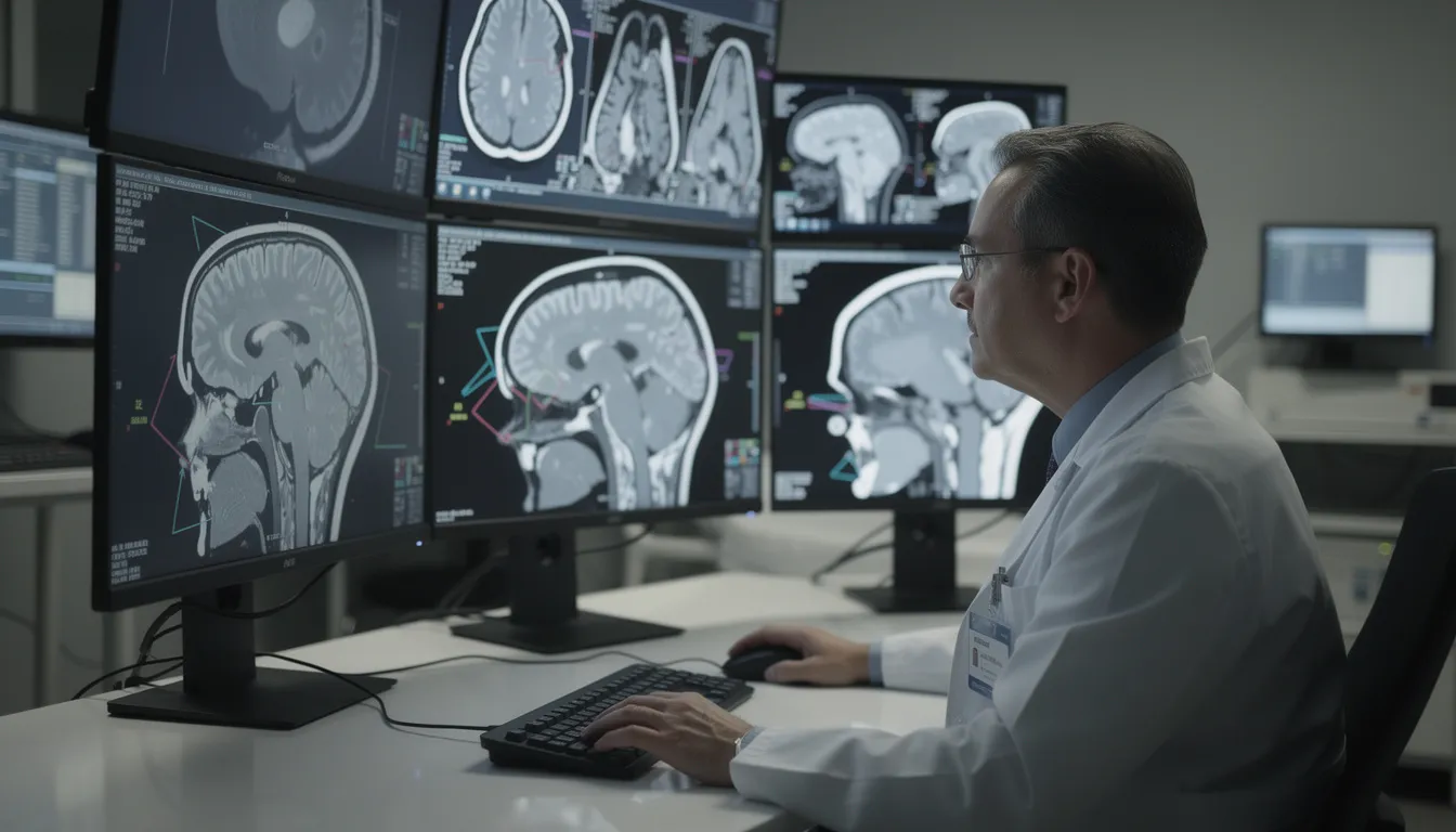
Interpreting mri results requires specialized training and expertise, as the detailed images provide complex information about tissue characteristics and anatomical relationships. Understanding the process helps patients know what to expect and how to prepare for follow-up care.
Radiologist analyzes images and sends detailed report to ordering physician within 24-48 hours in most cases. The radiologist examines multiple sequences and planes of imaging, looking for abnormalities in tissue signal characteristics, enhancement patterns, and anatomical structures.
Mri results are usually available within 24-48 hours through patient portals or direct communication from your healthcare provider. Urgent findings may be communicated immediately, while routine studies follow standard reporting timelines established by the facility.
Images produced show detailed soft tissue contrast not visible on conventional X-rays, providing information about:
- Tissue composition and characteristics
- Blood flow and vascular patterns
- Anatomical relationships between structures
- Presence of abnormal tissues or lesions
- Treatment response in ongoing conditions
Follow-up appointment scheduled to discuss findings and treatment recommendations based on the mri images and clinical context. Your physician will correlate the imaging findings with your symptoms and medical history to develop an appropriate treatment plan.
Additional imaging or tests may be recommended based on initial results if the MRI raises questions that require further investigation. Sometimes, different imaging methods or specialized MRI sequences provide complementary information for comprehensive diagnosis.
The quantitative mri measurements obtained during advanced examinations can track disease progression and treatment response over time. This longitudinal information proves valuable for managing chronic conditions and optimizing therapy effectiveness.
Magnetic resonance imaging represents one of medicine’s greatest technological achievements, providing physicians with unprecedented ability to see inside the human body without invasive procedures or ionizing radiation. From detecting early-stage cancers to mapping brain function before surgery, MRI continues to revolutionize medical diagnosis and treatment planning across virtually every medical specialty.
Understanding what an MRI is and how the process works empowers patients to approach their examinations with confidence and realistic expectations. Whether you’re scheduled for your first mri scan or helping a loved one prepare for their imaging test, this knowledge helps ensure the best possible experience and outcomes.
The safety profile, diagnostic capabilities, and patient comfort offered by modern mri technology make it an invaluable tool in contemporary healthcare. As imaging methods continue to advance, MRI will undoubtedly remain central to providing the detailed images physicians need to deliver optimal patient care.